Why are cells small?
Published: (June 8, 2026 at 03:10 PM EDT)
5 min read
Source: Hacker News
Source: Hacker News
A human body is built from 30 trillion cells — excluding microbes — that each arise from a lone, fertilized egg. These cells come in a multiplicity of shapes and sizes, with internal volumes spanning five orders of magnitude. The smallest human cell, a sperm, has a volume of just 30 µm³, whereas an oocyte has a volume of 4,000,000 µm³, making it the largest cell in the human body.1
What accounts for this huge range? A simplistic answer is that evolution has made each cell
the size best suited to its function. Maybe sperm are small because the body needs to make
many of them, and tiny cells cost less energy to make. (Sperm consist of little more than
DNA and a few mitochondria, which are necessary for providing energy to spin their whip-like
tails.) By contrast, an oocyte needs massive reserves of mitochondria and nutrients to
support early embryonic growth. In short, every cell is as large or small as it needs to be
— *within reason*.
But we can derive far more satisfying answers from physics.
The first major limit on a cell’s size is its surface area-to-volume
ratio. Assuming that a cell is roughly spherical in shape, its internal volume
grows proportionally to the *cube* of its radius, whereas its surface area grows
proportionally to the *square* of that radius. In other words, a cell’s
volume grows much faster than its surface area.
This ratio has big consequences for cell survival. The cell’s membrane funnels
nutrients into the cell and secretes waste. It’s also where the energy in a
prokaryotic cell — like *E. coli* — gets made. If the interior grows
too large relative to the membrane, the cell will not be able to produce enough energy or
excrete waste quickly enough to maintain all the ‘stuff’ inside, and metabolism
will slow down.
A second constraint is **diffusion**, or the tendency for molecules to
migrate from areas of high concentration to areas of lower concentration. This migration
dictates how quickly enzymes find substrates, or how signaling molecules reach receptors,
and how often ribosomes collide with messenger RNAs. Inside a cell, nearly everything
happens by chance encounters amongst molecules! As a cell’s volume grows, though,
the chance that these encounters will happen decreases (assuming the total numbers of
molecules stay constant).
A molecule’s diffusion rate changes based on various factors. The cytoplasm
is extremely crowded, for example, and so molecules spend lots of time ricocheting off obstacles,
delaying their arrival at a distant location. Every protein in a cell collides with about
10 billion water molecules per second on average. The vast majority of proteins in a bacterium have diffusion coefficients of only 5 to 10 µm2
per second (a measure of how quickly molecules spread through space). Some molecules also aggregate or stick to charged surfaces, further slowing
their movement.[2](#fn2)
In general, large molecules diffuse slower than small ones.
Metabolites in *E. coli* can diffuse from one side of the cell to the other in
milliseconds, which means collisions — and cellular *outcomes* — happen
quickly. A typical protein takes just
[0.01 seconds](https://bionumbers.hms.harvard.edu/bionumber.aspx?id=103801)
to traverse a bacterium’s diameter (about 1 micrometer), but the same
protein would take around four minutes to move one millimeter and more than six
hours to move one centimeter. This is, in part, why cells are so tiny.
With these constraints in mind, we can begin to speculate as to why various cells are
shaped the way they are.
Red blood cells are tiny and shaped like biconcave discs to aid with diffusion; by
abandoning a spherical shape and evolving more toward a ‘donut,’ they increase
their surface area without compromising their compact volume. This, in turn, enhances their
ability to exchange oxygen with cells in the body. Their small size (just 8 micrometers
across) also helps them move through narrow capillaries.
In contrast, oocytes can grow so large (around 100 micrometers in diameter), in part,
because they are less metabolically active than other types of human cells — and thus
don’t depend so much on random collisions. They stockpile nutrients
during oogenesis to wait out fertilization. Eukaryotic cells also grow large,
in general, because they’ve evolved *compartmentalization*; by modularizing
specific functions into organelles, they bring molecules closer together to help get the
job done.
Cell sizes are not fixed, however, even within a single species. Cells often swell as they
increase their production of proteins and metabolites in preparation for division. This is
in line with biology’s only rule: namely, there are exceptions to every rule!
Case in point: a giant bacterium called *Thiomargarita magnifica* can extend about
one centimeter in length, so large that it can be seen by the naked eye. It does so by
breaking the surface area-to-volume rule, filling between 65–80 percent of its
internal volume with an empty vacuole. In other words, it pushes most of its
molecules to the cell periphery, thus shortening diffusion
distances.[3](#fn3)
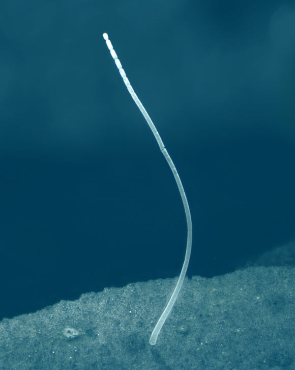
*Thiomargarita magnifica* is a bacterial species that can extend about one centimeter in length, several orders of magnitude more than *E. coli*. These microbes are visible to the naked eye. Credit: Jean-Marie Volland
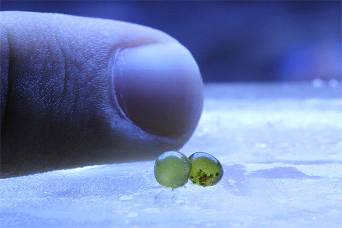
Bubble algae (aka *Valonia ventricosa*). Credit: Trident's Cove
Despite their variety, these architectures still hinge on molecules bumping into each
other, guided by the immutable laws of physics. Or, as D’Arcy Wentworth Thompson
mused in *On Growth and Form* (1917), “The form of an object is a
‘diagram of forces.’” Cells bear witness to both internal and external
forces; they are constrained by diffusion and shaped by the delicate trade-off between
volume and surface area.